이번 호에서는 WHO 2008 분류 중 골수증식종양(myeloproliferative neoplasm)에
속하는 진성적혈구증가증(polycythemia vera, PV)과 진성혈소판증가증(essential thrombocythemia,
ET) 진단의 최신지견에 대해 정리하였습니다.
1. 개요
1) PV 및 ET의 WHO 분류 및 특징
- 골수성 혈액종양의 5대 분류 중 골수증식종양(myeloproliferative neoplasm, MPN)에 속함 (Table
1 참고)
- 편의적으로 PV, ET, 일차골수섬유증(primary myelofibrosis, PMF)을 “BCR-ABL1-negative
MPN”으로 분류하며 줄기세포의 클론성 증식이 특징임
2) PV 및 ET의 분자유전학적 특징
- 질병유발원인 돌연변이에 대해서는 아직 밝혀지지 않음
- 관련 돌연변이로는JAK2 (Janus kinase 2; 9p24) 및 MPL
(myeloproliferative leukemia virus
oncogene; 1p34) 돌연변이가 알려짐(이들 돌연변이는 다른 골수성 혈액종양에서 관찰되기도
하고, TET2나 IDH 돌연변이와 같은 다른 돌연변이가 MPN에서
관찰되기도 함)
- 각 환자군에서의 돌연변이 관찰빈도
■ JAK2 V617F 양성 또는 allele burden의 증가: PV나
ET환자의 생존율이나 백혈병으로의 전환에 영향을 미치지는 않음. 일반적으로 고령, 높은 혈색소수치,
백혈구증가증, 혈소판감소증과 관련이 있음 • JAK2 V617F
양성 ET: 동맥혈전증의 위험도 높음. 진성혈소판증가증 후 골수섬유화 (post-ET MF)로의 진행
위험도 낮음 • JAK2 V617F 양성 PV: mutant allele burden이 높을수록
가려움증 및 섬유증 전환의 위험도 높음
■ JAK2 exon 12 돌연변이: JAK2 V617F와 유사한 예후를 보이지만, 진단 시 골수에서
세 계열 중 주로 적혈구계 세포증식, 혈청 적혈구형성인자(erythropoietin, Epo) 수치
저하, 더 젊은 연령을 보이는 것으로 보고됨
■ MPL 돌연변이: 일관되지는 않으나 고령, 여성, 낮은 혈색소수치, 높은 혈소판수치와 관련이 있으며,
생존율이나 백혈병으로의 전환과는 관련이 없는 것으로 보고됨
Table 1. WHO classification of myeloid malignancies
1. Acute myeloid leukemia (AML) and related precursor
neoplasmsa
5. Myeloid and lymphoid neoplasms with eosinophilia
and abnormalities of PDGFRA,cPDGFRB,c
or FGFR1c 5.1.
Myeloid and lymphoid neoplasms with PDGFRA rearrangement 5.2.
Myeloid neoplasms with PDGFRB rearrangement 5.3.
Myeloid and lymphoid neoplasms with FGFR1 abnormalities
aAcute myeloid leukemia-related precursor
neoplasms include “therapy-related myelodysplastic syndrome” and “myeloid
sarcoma”. bEither mono- or bi-cytopenia: hemoglobin level
<10 g/dL, absolute neutrophil count <1.8 X 10⁹/L, or platelet
count <100 X 109/L. However, higher blood counts do not exclude
the diagnosis in the presence of unequivocal histological/cytogenetic evidence
for myelodysplastic syndrome. cGenetic rearrangements involving
platelet-derived growth factor receptor α/β (PDGFRA/PDGFRB)
or fibroblast growth factor receptor 1 (FGFR1).
2. 진단
1) PV와 ET의 진단
- 임상적 그리고 검사의 특성을 고려한 WHO 진단기준을 바탕으로 함(Table 2 참고)
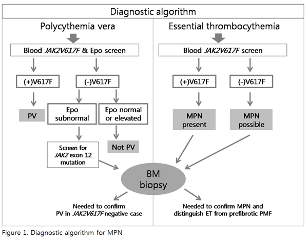
- 말초혈액의 JAK2 V617F 검사를 시작으로 하는 진단 알고리듬은 Figure 1 참고
2) PV의 진단
- JAK2 V617F 검사는 매우 민감하며(민감도 97%) 다른 원인의 적혈구용적률(hematocrit) 증가와
PV를 거의 100% 감별해 낼 수 있음
- 혈청 적혈구형성인자는 PV 환자의 85% 이상에서 저하된 수치를 보이기 때문에 동시에 측정하게 되는
경우 JAK2 V617F 위양성이나 위음성의 위험성을 줄일 수 있음
- JAK2 V617F 음성이면서 저하된 적혈구형성인자 수치를 보이는 경우 약 3%를 차지하는 것으로 알려진
JAK2 V617F-음성 PV 환자를 감별하기 위해 JAK2 exon 12 돌연변이 검사 시행이
필요함
- PV 진단을 위해서 골수검사가 필수적이지는 않음
■ JAK2 V617F 양성: MPN이 있는 것으로 확진 가능
■ JAK2 V617F 음성: ET환자의 40%는 JAK2 V617F 음성일 수 있기 때문에 ET를
배제할 수 없음
- 다른 JAK2 V617F 양성 MPN (또는 MDS/MPN)의 양상이 ET와
유사할 수 있으므로 정확한 진단을 위해서 골수 검사가 필요함
■ Prefibrotic PMF
•거핵구의 비정상적인 성숙 형태, 핵의 과다염색성과 불규칙하게 접히는 모양을 보임
cf) ET의 거핵구는 크고 성숙된 형태로 보임 • 최근 대규모 국제연구에서 ET와 pre-fibrotic
PMF를 형태학적으로 정확하게 감별하는 것이 임상적 예후를 판정하는데 중요함을 보고함1
■ Refractory anemia with ring sideroblasts with marked
thrombocytosis (RARS-T)
- JAK2 V617F 음성인 경우 CML과의 감별
■ CML: BCR-ABL1 돌연변이 검사 또는 작고 저분엽 형태의 거핵구
관찰로 ET와 쉽게 감별 가능
4) Post-PV 또는 post-ET MF의 진단: International Working
Group for MPN Research and
Treatment (IWG-MRT)의 진단 기준을 따름(Table 3 참고)
Table 2. World Health Organization (WHO) Diagnostic
Criteria for Polycythemia Vera, Essential Thrombocythemia and Primary Myelofibrosis
2008 WHO Diagnostic Criteria
Polycythemia veraa
Essential thrombocythemiaa
Primary myelofibrosisa
Major criteria
Hgb >18.5 g/dL (men), >16.5 g/dL (women),
orb
Presence of JAK2 V617F or JAK2 exon 12 mutations
Platelet count ≥450 x 109/L
Megakaryocyte proliferation with large and mature morphology
Not meeting WHO criteria for CML, PV, PMF, MDS, or other myeloid
neoplasm
Demonstration of JAK2 V617F or other clonal marker
or no evidence of reactive thrombocytosis
Megakaryocyte proliferation and atypiac accompanied
by either reticulin and/or collagen fibrosis, ord
Not meeting WHO criteria for CML, PV, MDS, or other myeloid
neoplasm
Demonstration of JAK2 V617F or other clonal marker
or no evidence of reactive marrow fibrosis
Minor criteria
BM trilineage myeloproliferation
Subnormal serum Epo level
EEC growth
Leukoerythroblastosis
Increased serum LDH level
Anemia
Palpable splenomegaly
BM, bone marrow; Hgb, hemoglobin; Hct, hematocrit;
Epo, erythropoietin; EEC, endogenous erythroid colony; WHO, World Health
Organization; CML, chronic myelogenous leukemia; PV, polycythemia vera;
PMF, primary myelofibrosis; MDS, myelodysplastic syndromes; LDH, lactate
dehydrogenase. aPV diagnosis requires meeting
either both major criteria and 1 minor criterion or the
first major criterion and 2 minor criteria. ET diagnosis
requires meeting all 4 major criteria. PMF diagnosis
requires meeting all 3 major criteria and 2 minor criteria. bor
Hgb or Hct >99th percentile of reference range for age, sex, or altitude
of residence or red cell mass >25% above mean normal predicted
value or Hgb >17 g/dL (men)/>15 g/dL (women) if associated
with a sustained increase of ≥2 g/dL from baseline that cannot be attributed
to correction of iron deficiency. cSmall to large megakaryocytes
with aberrant nuclear/cytoplasmic ratio and hyperchromatic and irregularly
folded nuclei and dense clustering. dor In the absence of
reticulin fibrosis, the megakaryocyte changes must be accompanied by increased
marrow cellularity, granulocytic proliferation, and often decreased erythropoiesis
(i.e., pre-fibrotic PMF).
Table 3. International Working Group for Myeloproliferative
Neoplasms Research and Treatment (IWG-MRT) Recommended Criteria for Post-Polycythemia
Vera and Post-Essential Thrombocythemia Myelofibrosis
Criteria for post-polycythemia vera myelofibrosis
Required criteria:
Documentation of a previous diagnosis of polycythemia vera
as defined by the WHO criteria (see Table 2)
Bone marrow fibrosis grade 2-3 (on 0-3 scale) or grade 3-4
(on 0-4 scale) (see footnote for details)
Additional criteria (2 are required):
Anemia or sustained loss of requirement for phlebotomy in the
absence of cytoreductive therapy
A leukoerythroblastic peripheral blood picture
Increasing splenomegaly defined as either an increase in palpable
splenomegaly of ≥5cm (distance of the tip of the spleen from the
left costal margin) or the appearance of a newly palpable splenomegaly
Development of ≥1 of 3 constitutional symptoms: >10% weight
loss in 6 months, night sweats, unexplained fever (>37.5°C)
Criteria for post-essential thrombocythemia
myelofibrosis
Required criteria:
Documentation of a previous diagnosis of essential thrombocythemia
as defined by the WHO criteria (see Table 2)
Bone marrow fibrosis grade 2-3 (on 0-3 scale) or grade 3-4
(on 0-4 scale) (see footnote for details)
Additional criteria (2 are required):
Anemia and a ≥2 g/dL decrease from baseline hemoglobin level
A leukoerythroblastic peripheral blood picture
Increasing splenomegaly defined as either an increase in palpable
splenomegaly of ≥5 cm (distance of the tip of the spleen from
the left costal margin) or the appearance of a newly palpable
splenomegaly
Increased lactate dehydrogenase
Development of ≥1 of 3 constitutional symptoms: >10% weight
loss in 6 months, night sweats, unexplained fever (>37.5°C)
Grade 2-3 according to the European classification:
diffuse, often coarse fiber network with no evidence of collagenization
(negative trichrome stain) or diffuse, coarse fiber network with areas of
collagenization (positive trichrome stain).
Grade 3-4 according to the standard classification: diffuse and dense increase
in reticulin with extensive intersections, occasionally with only focal
bundles of collagen and/or focal osteosclerosis or diffuse and dense increase
in reticulin with extensive intersections with coarse bundles of collagen,
often associated with significant osteosclerosis.
3. 참고문헌
Barbui T, Thiele J, Passamonti F, Rumi F, Boveri E, Ruggeri M, et
al. Survival and disease progression in essential thrombocythemia are
significantly influenced by accurate morphologic diagnosis: an international
study. J Clin Oncol 2011;29:3179-84.
(http://www.ncbi.nlm.nih.gov/pubmed/21747083)
Tefferi A. Polycythemia vera and essential thrombocythemia: 2012
update on diagnosis, risk stratification, and management. Am J Hematol
2012;87:285-93.
(http://www.ncbi.nlm.nih.gov/pubmed/22331582)